 RadiologyQuizLab
CROSS-SECTIONAL ANATOMY · CT & MRI
Cross-sectional anatomy
Cross-sectional anatomy — often just called sectional anatomy — is how you actually read a CT or MRI — slice by slice, plane by plane. Unlike the drawn diagrams in a textbook, real imaging shows structures as shifting shades of grey that change with the modality and the level, so the only way to get fluent is to name them on real images. This page explains the three planes, how CT and MRI differ, and the landmarks to learn first — then lets you practise the exact skill, free.
Try it now — read the slice, name the arrowed structure:
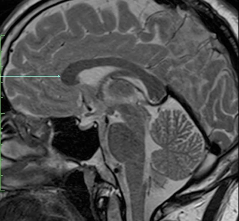
Which anatomical structure is arrowed?
Single answer — type the structure
Correct — Genu of corpus callosum
The three planes
Every cross-sectional study is built from three orthogonal planes. The axial (transverse) plane cuts the body head-to-toe into slices and is the workhorse of CT. The coronal plane divides front from back, seen as if looking at a standing person face-on; the sagittal plane divides left from right, and the midline sagittal is where structures such as the corpus callosum and brainstem are best appreciated. Modern scanners acquire a volume that can be reformatted into any plane, so the same structure can be checked from three angles — learning to picture it across all three is the core skill of radiological anatomy.
CT vs MRI
CT and MRI both produce cross-sectional images but show tissue completely differently. CT measures X-ray density in Hounsfield units — bone is bright, air is black, soft tissues sit in between — which makes it fast and excellent for bone, lung, acute bleeding and trauma; iodinated contrast then lights up vessels and organs in phases. MRI instead maps the behaviour of water protons across many sequences (T1, T2, FLAIR, diffusion and more), giving far superior soft-tissue contrast, which is why brain, spine, joint and pelvic anatomy are usually learned on MRI. The same structure looks different on each — recognising it either way is exactly what practice builds.
What to learn first
Start with the landmarks that orient every study, then let the smaller structures fall into place around them. On a chest CT that means the great vessels — aorta, pulmonary arteries and the venae cavae — and the four heart chambers; on an abdominal CT, the liver segments defined by the hepatic and portal veins, the pancreas against the splenic vein, and the kidneys with their vessels; on a brain MRI, the ventricles, basal ganglia, thalami and brainstem. Once these are automatic you can read outward to the muscles, nerves and finer vessels, and switch confidently between CT and MRI of the same region.
Keep practising by region
Work through anatomy one modality and region at a time — brain MRI, abdominal CT, cardiac MRI and more — and let each explanation fix the structure in memory.
Ready for the full set?
Get 20 free questions across 8 body regions — name the structure, get the explanation, and track what sticks.
Start free — 20 questions