 RadiologyQuizLab
CARDIAC MRI ANATOMY · SSFP CINE
Cardiac MRI anatomy
Cardiac MRI anatomy is learned through its standard planes. Practise naming the structures on real cardiac MRI images — the fastest way to make cardiac anatomy stick. Free, no account needed.
Try it now — read the slice, name the arrowed structure:
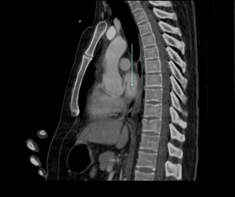
Which anatomical structure is arrowed?
Single answer — type the structure
Correct — Left atrium
The cardiac planes
Cardiac MRI uses dedicated planes — two-, three- and four-chamber, and the short axis — rather than pure body planes. Learning to recognise each view is the first step.
Chambers, valves, vessels
Identify the four chambers, the AV and semilunar valves, and the great vessels as they come and go across the planes. The short-axis stack is where the ventricles and walls are assessed.
Why cine and SSFP
Bright-blood SSFP cine gives the crisp blood–myocardium border cardiac anatomy relies on. Recognising structures on these sequences is what practice builds.
Keep practising by region
Move through the rest of cross-sectional anatomy one region at a time — brain and abdominal CT, cardiac MRI and more — and let each explanation fix the structure in memory.
Ready for the full set?
Get 20 free questions across 8 body regions — name the structure, get the explanation, and track what sticks.
Start free — 20 questions