 RadiologyQuizLab
MRI ANATOMY · T1 · T2 · FLAIR
MRI anatomy
MRI anatomy is best learned on real images, naming the structure yourself. Practise across sequences and regions — the fastest way to make imaging anatomy stick. Free, no account needed.
Try it now — read the slice, name the arrowed structure:
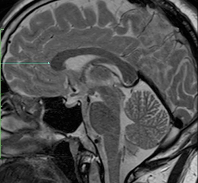
Which anatomical structure is arrowed?
Single answer — type the structure
Correct — Genu of corpus callosum
Reading the sequences
T1, T2, FLAIR, diffusion and more each show tissue differently — the anatomy stays the same, only the signal changes. Knowing your sequence is the first step to naming what you see.
Region by region
Brain, spine, joints and pelvis each reward MRI's soft-tissue detail. Start from the obvious landmarks and let the smaller structures fall into place around them.
MRI vs CT
MRI leads on soft tissue and multiplanar detail; CT is faster and better for bone and acute blood. Recognising a structure on either is what practice builds.
Keep practising by region
Move through the rest of cross-sectional anatomy one region at a time — brain and abdominal CT, cardiac MRI and more — and let each explanation fix the structure in memory.
Ready for the full set?
Get 20 free questions across 8 body regions — name the structure, get the explanation, and track what sticks.
Start free — 20 questions