 RadiologyQuizLab
BRAIN MRI ANATOMY · T1 · T2 · FLAIR
Brain MRI anatomy
Brain MRI anatomy is best learned the way you'll use it: on real slices, naming the structure yourself. Practise on axial, coronal and sagittal images across the main sequences — the quickest way to make neuroanatomy stick. Free, no account needed.
Try it now — read the slice, name the arrowed structure:
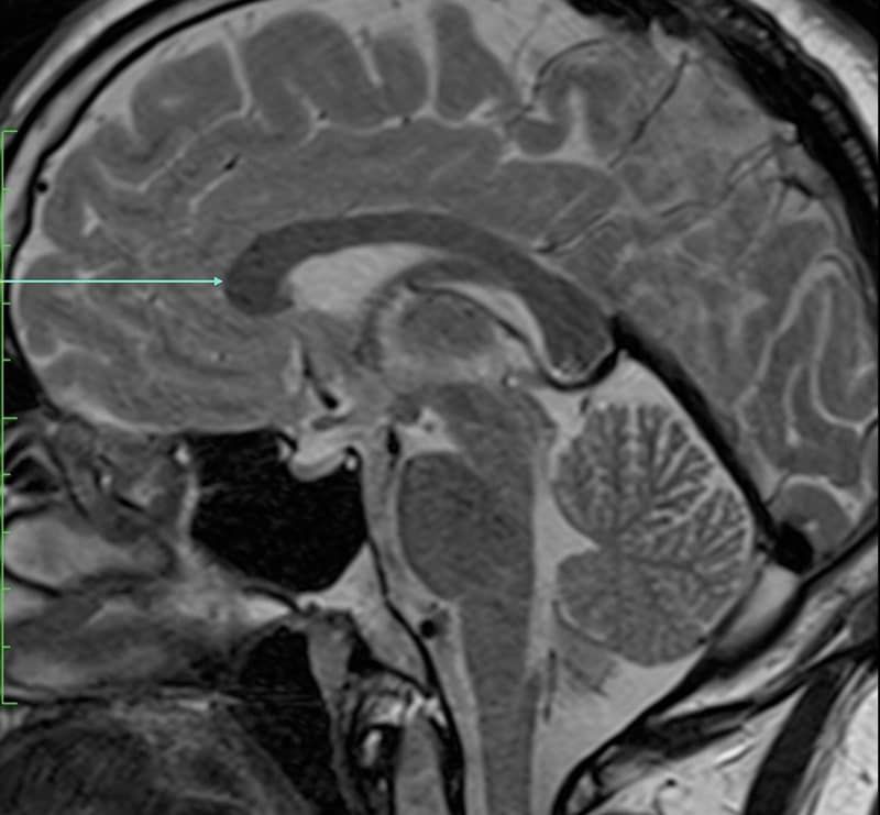
Which anatomical structure is arrowed?
Single answer — type the structure
Correct — Genu of corpus callosum
Reading the sequences
The same brain looks different on each MRI sequence. On T1 the CSF is dark and white matter is bright — good for anatomy. On T2 and FLAIR the CSF is bright (suppressed on FLAIR), which sharpens the ventricles and cortex. Knowing which sequence you're on is the first step to naming what you see; the anatomy underneath never changes, only its signal.
The landmarks that orient you
Start from the midline sagittal: the corpus callosum, brainstem (midbrain, pons, medulla), cerebellum and pituitary stand out clearly. On axial slices, work outward from the ventricles — lateral, third and fourth — to the basal ganglia (caudate, putamen, globus pallidus), thalami and internal capsule. These few structures anchor almost every study.
Cortex, deep grey and vessels
Once the core landmarks are automatic, add the lobes and key gyri (pre- and post-central), the hippocampi on coronal views, and the main vessels of the circle of Willis on flow-sensitive sequences. Naming them repeatedly on real images is what turns recognition into recall.
Keep practising by region
Move through the rest of cross-sectional anatomy one region at a time — abdominal CT, cardiac MRI and more — and let each explanation fix the structure in memory.
Ready for the full set?
Get 20 free questions across 8 body regions — name the structure, get the explanation, and track what sticks.
Start free — 20 questions