 RadiologyQuizLab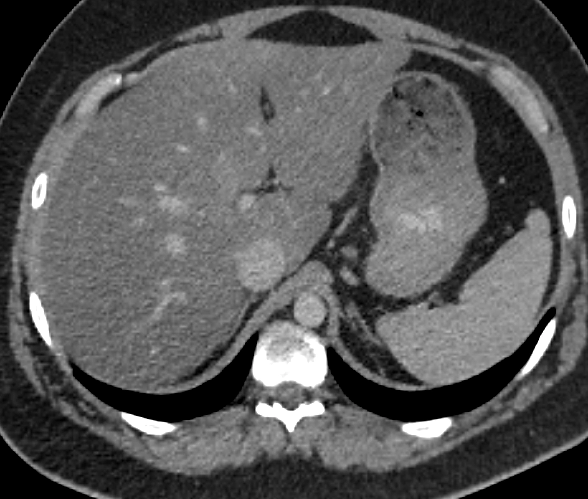
How to use this CT anatomy atlas
- Select the liver or a kidney from the structure list. The crosshair moves to a point inside that organ in all three slices.
- Drag the 3D model to understand the organ’s position. Click a visible organ to move the crosshair to the point you selected.
- Move a slice slider to follow a structure through the scan. Click another point in an image to update the other views.
- Hide selected models to see deeper structures. Then practise naming structures from memory in the anatomy quizzes.
Understand axial, coronal and sagittal CT images
An axial image cuts across the body; a coronal image separates front and back; a sagittal image separates left and right. These views describe the same volume from different directions. The shared crosshair helps you connect a small circular structure on one slice with a longer structure in another plane.
Why is the patient’s right on the left of the image?
Axial CT images here are displayed as if you were looking up from the patient’s feet. The R and L markers refer to the patient, so the liver appears on the screen’s left. Coronal views face the patient. On the sagittal view, anterior is on the left and posterior on the right. A/P mean anterior/posterior; S/I mean superior/inferior.
From a 3D atlas to active recall
Use the atlas to understand spatial relationships before testing yourself. Follow the kidneys on either side of the spine and locate the aorta and inferior vena cava. The atlas uses a fixed soft-tissue window (width 400, level 50) and eight segmented structures. It is a focused abdominal learning tool, not a complete anatomical atlas or a viewer for uploading your own scans.
Try 20 free anatomy questionsQuestions about the explorer
Is this CT anatomy explorer free?
Yes. The atlas works without an account. The separate RadiologyQuizLab quiz app offers 20 free questions after registration.
Are these real CT images?
Yes. The slices and 3D surfaces come from the same TotalSegmentator CT scan and segmentation masks. The images are resampled and the surfaces simplified for the browser; fine details may be reduced.
Can I use it on my phone?
Yes. The views stack vertically on a small screen. Use the organ list and sliders to navigate, and pinch the 3D model to zoom. The 3D view requires WebGL.
Continue learning radiological anatomy
Can you name it without the label?
Practise identifying structures on real medical images. Type your answer, check it and revisit the structures you missed.
Try 20 free anatomy questions