 RadiologyQuizLab
FRCR PART 1 · ANATOMY MODULE
FRCR Part 1 Anatomy
The anatomy module of the First FRCR is an image-viewing exam: you're shown a radiological image with a structure marked, and you name it. This guide explains how the exam works, what's tested and how to revise — then lets you practise the same skill on real images, free.
The exam skill, in one question — read the image, name the marked structure:
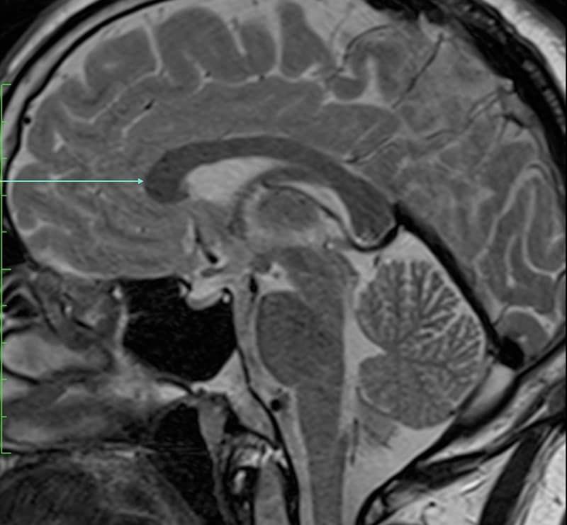
Which anatomical structure is arrowed?
Single answer — type the structure
Correct — Genu of corpus callosum
What the anatomy exam actually is
The First FRCR (CR1) has two modules — anatomy and physics — sat on separate days at a test centre. The anatomy module is a viewing paper: a series of images, each with an arrow or marker on one structure, and you type the name of that structure. There are no multiple-choice options — it's pure recall, which is exactly how you read images on the job.
Exam format at a glance
Expect around a hundred marked structures across every modality — plain radiographs, fluoroscopy, ultrasound, CT, MRI and nuclear medicine — and every body region. Answers are short free text and each structure is marked individually. Exact question counts, pass mark and exam dates change each sitting, so always confirm the current details on the official Royal College of Radiologists exam page.
Official exam details — Royal College of Radiologists →
What's tested
Everything: cross-sectional anatomy on CT and MRI, projectional anatomy on plain films, vascular anatomy on angiography, and the anatomy behind ultrasound and fluoroscopy studies. Neuro, head and neck, chest, abdomen and pelvis, and musculoskeletal all appear. The exam rewards recognising the same structure across modalities and planes.
How to revise (what works)
Start early and study region by region — bones, then vessels, then the soft tissues of each area. Learn on real images, not diagrams, because the exam uses real images. Use active recall: cover the label, name the structure, then check — repetition on real cases is what turns recognition into instant recall. Ryan's Anatomy for Diagnostic Imaging is the usual companion alongside daily image practice.
FRCR Part 1 anatomy: common questions
What is the FRCR Part 1 anatomy pass mark?
The pass mark is set for each sitting by the RCR and isn't a fixed number — check the current figure on the official RCR exam page. Consistent daily practice matters more than chasing the exact threshold.
When are the FRCR Part 1 anatomy exam dates?
Dates and booking deadlines are published each year by the RCR. Confirm the current sitting on the RCR website before you plan your revision.
Are there FRCR anatomy mock papers or practice questions?
Yes — practising on marked images is the single most useful preparation. You can practise the exact name-the-structure format free here, on real CT, MRI and radiograph images, no account needed.
What's the best resource for FRCR anatomy?
Combine a reference atlas (Ryan's Anatomy for Diagnostic Imaging is the usual one) with daily image practice. Reading real images and naming structures beats passively reading a book.
Ready to practise like the exam?
Get 20 free questions across 8 body regions — name the structure, get the explanation, and track what sticks. No card needed.
Practise free — 20 questions