 RadiologyQuizLab
KNEE MRI ANATOMY · SAGITTAL · CORONAL
Knee MRI anatomy
Knee MRI is one of the most-requested musculoskeletal studies, and its anatomy is dense: menisci, ligaments, cartilage and tendons all packed into a small joint and spread across several planes. The only reliable way to learn it is to name each structure on real sagittal, coronal and axial images. This guide walks through the menisci and cartilage, the ligaments, and the extensor mechanism — then lets you practise the exact skill, free.
Try it now — read the slice, name the arrowed structure:
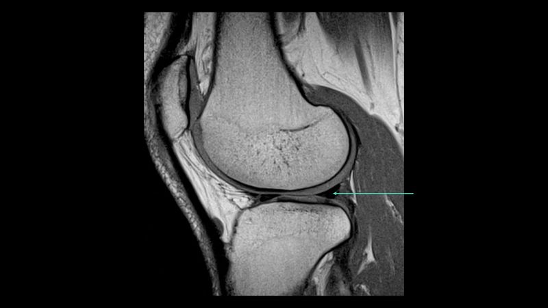
Which anatomical structure is arrowed?
Single answer — type the structure
Correct — Posterior horn of lateral meniscus
Menisci and cartilage
Start with the medial and lateral menisci: on sagittal images they appear as two dark triangles — the anterior and posterior horns — that should be smooth and uniformly low in signal. Follow them onto the coronal plane, where the body of each meniscus and the meniscal roots are seen. Then assess the articular cartilage over the femoral condyles, the tibial plateau and behind the patella, judging its thickness and continuity. These are the structures most knee studies turn on, so getting fluent with their normal appearance comes first.
Ligaments
The cruciate ligaments are read on the sagittal plane: the anterior cruciate runs obliquely from the tibia up to the lateral femoral condyle and is normally taut and straight, while the posterior cruciate forms a smooth dark arc behind it. The collateral ligaments are best seen on coronals — the medial collateral as a stripe along the inner joint line, the lateral (fibular) collateral running to the fibular head with the biceps femoris. Knowing each ligament's normal course and signal is the key to reading the knee with confidence.
Tendons and the extensor mechanism
Complete the study with the extensor mechanism — the quadriceps tendon inserting on the patella and the patellar tendon running down to the tibial tuberosity — plus the popliteus and the tendons around the joint. Behind the knee, identify the popliteal vessels and the neurovascular bundle. Naming these on real images, over and over, is what turns slow recognition into the instant recall you need when reading a case or sitting an exam.
Keep practising by region
Move through the rest of imaging anatomy one region at a time — brain and body CT, cardiac and musculoskeletal MRI — and let each explanation fix the structure in memory.
Ready for the full set?
Get 20 free questions across 8 body regions — name the structure, get the explanation, and track what sticks.
Start free — 20 questions