 RadiologyQuizLab
CT ANATOMY · AXIAL · CORONAL · SAGITTAL
CT anatomy
CT anatomy is read slice by slice and region by region, and the method barely changes as you move down the body. This hub explains how a CT dataset is built, how to work through it one region at a time, and the role of contrast — then links to focused quizzes for the head, chest, abdomen and pelvis so you can practise on real images, free.
Try it now — read the slice, name the arrowed structure:
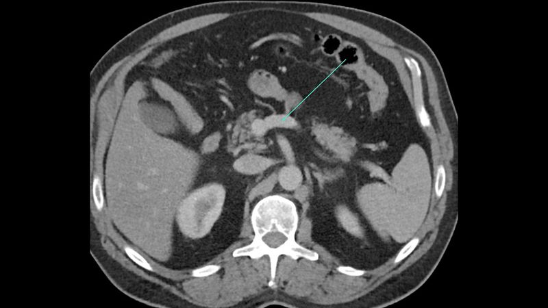
Which anatomical structure is arrowed?
Single answer — type the structure
Correct — Splenic vein
How a CT is built
A CT scanner acquires a stack of thin axial slices that can be reformatted into coronal and sagittal planes, so a single study can be reviewed from any angle. Each pixel carries a density value in Hounsfield units — about −1000 for air, 0 for water, and several hundred to over a thousand for bone — and the window you choose decides what stands out: a bone window for the skeleton, a soft-tissue window for organs, a lung window for the airways and parenchyma. This is what lets you read the same slice three different ways.
Region by region
Head, chest, abdomen and pelvis each have their own landmarks, but the approach is identical: find the great vessels and the bones first, then place the organs around them. In the chest that means the aorta, pulmonary arteries and heart; in the abdomen, the aorta and IVC with the liver, pancreas and kidneys; in the pelvis, the iliac vessels with the bladder and bowel. Once the fixed landmarks are automatic, the smaller structures of each region fall into place.
The role of contrast
Intravenous contrast changes a CT completely. In the arterial phase the aorta and its branches light up; in the portal-venous phase the solid organs enhance most and the veins opacify; later phases show the collecting systems of the kidneys. Knowing which phase you are looking at tells you what should be bright and helps you name vessels and organs correctly. Without contrast, bone and acute blood dominate — which is exactly what you want in trauma.
Keep practising by region
Move through the rest of cross-sectional anatomy one region at a time — brain and abdominal CT, cardiac MRI and more — and let each explanation fix the structure in memory.
Ready for the full set?
Get 20 free questions across 8 body regions — name the structure, get the explanation, and track what sticks.
Start free — 20 questions