 RadiologyQuizLab
CT ABDOMEN ANATOMY · AXIAL CT
CT abdomen anatomy
CT abdomen anatomy is the backbone of body imaging. Practise naming the structures on real axial CT slices — the quickest way to make abdominal anatomy stick. Free, no account needed.
Try it now — read the slice, name the arrowed structure:
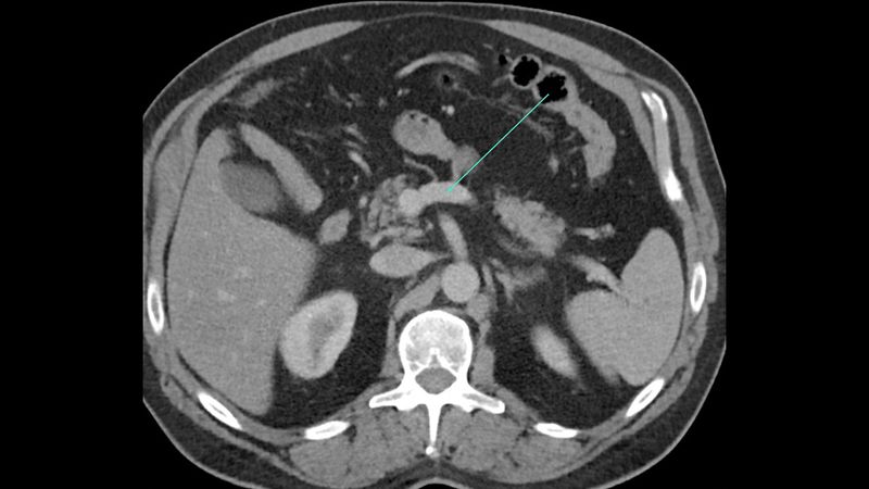
Which anatomical structure is arrowed?
Single answer — type the structure
Correct — Splenic vein
Orient yourself first
Start from the great vessels — aorta and IVC — and the vertebral level, then place the solid organs around them. Reading top-to-bottom, the liver and spleen give way to the kidneys, then bowel and pelvis.
Liver, pancreas, kidneys
Identify the liver segments around the hepatic and portal veins, the pancreatic head–body–tail against the splenic vein, and the kidneys with their vessels and collecting systems. These are the structures examiners return to.
The role of contrast
IV contrast phases (arterial, portal venous, delayed) change what stands out — vessels, organ enhancement, then the collecting system. Knowing the phase helps you name what you see.
Keep practising by region
Move through the rest of cross-sectional anatomy one region at a time — brain and abdominal CT, cardiac MRI and more — and let each explanation fix the structure in memory.
Ready for the full set?
Get 20 free questions across 8 body regions — name the structure, get the explanation, and track what sticks.
Start free — 20 questions