 RadiologyQuizLab
CT CHEST ANATOMY · AXIAL CT
CT chest anatomy
CT chest anatomy is where mediastinal and vascular landmarks come together. Practise naming the structures on real axial CT slices — the fastest way to make thoracic anatomy stick. Free, no account needed.
Try it now — read the slice, name the arrowed structure:
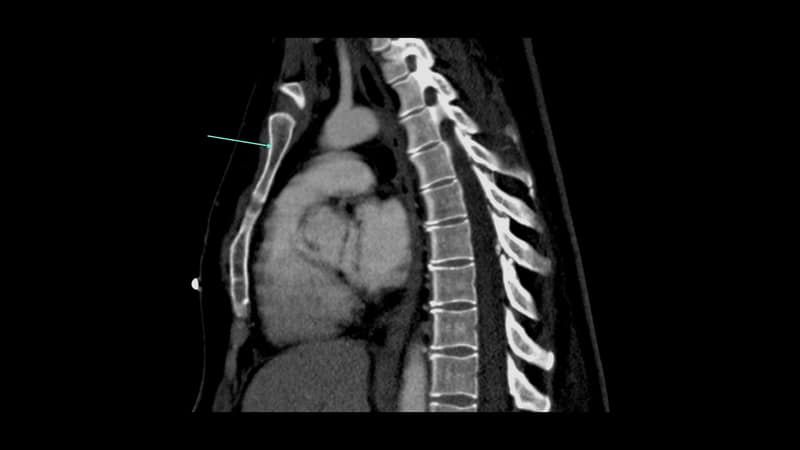
Which anatomical structure is arrowed?
Single answer — type the structure
Correct — Manubrium of sternum
Mediastinum and vessels
Anchor on the great vessels — aortic arch and its branches, pulmonary arteries, SVC — then place the heart chambers and the airway. These landmarks orient every chest CT.
Lung windows and airways
Switch to lung windows to follow the trachea, carina and bronchi into the lobes and segments. The fissures divide the lungs into the segments you'll be asked to name.
Windows change everything
The same slice reads differently on mediastinal, lung and bone windows. Recognising a structure across windows is the core skill of chest CT anatomy.
Keep practising by region
Move through the rest of cross-sectional anatomy one region at a time — brain and abdominal CT, cardiac MRI and more — and let each explanation fix the structure in memory.
Ready for the full set?
Get 20 free questions across 8 body regions — name the structure, get the explanation, and track what sticks.
Start free — 20 questions