 RadiologyQuizLab
CT HEAD ANATOMY · AXIAL CT
CT head anatomy
CT head anatomy is the first thing you read in any emergency scan, and the structures come thick and fast. Because a head CT is reviewed on several windows and the same slice means different things on each, fluency comes only from naming structures on real images. This guide covers how to read a head CT, the landmarks that orient every study, and how CT compares with MRI — then lets you practise on real axial slices, free.
Try it now — read the slice, name the arrowed structure:
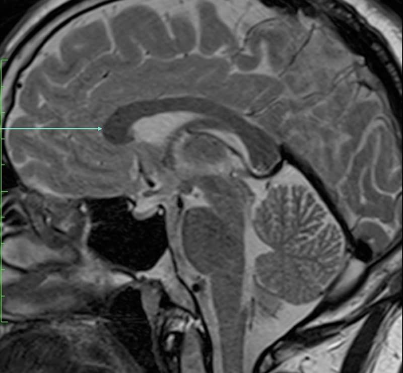
Which anatomical structure is arrowed?
Single answer — type the structure
Correct — Genu of corpus callosum
Reading a head CT
A head CT is read on a few standard windows. The brain window narrows the greyscale so grey and white matter separate, revealing the ventricles, basal ganglia and cortex; the bone window widens it so the skull vault, skull base and sinuses stand out; a subdural (blood) window helps pick up thin collections along the falx and tentorium. The raw data is identical — only the window changes — so learning which window shows which structure is half the battle. Work systematically from the vertex down to the skull base.
The landmarks to know
Orient yourself on the ventricles first — the lateral ventricles with their frontal and occipital horns, then the third and fourth ventricles on the midline. Around them sit the basal ganglia (caudate, putamen, globus pallidus), the thalami and the internal capsule, which make up the deep grey and white matter you are asked to name most often. At the skull base, identify the sphenoid and petrous temporal bones, the sella and the major foramina. These few landmarks anchor almost every head CT.
CT vs MRI of the brain
CT is fast, universally available and superb for bone, acute blood and calcification, which is why it is first-line in trauma and suspected stroke. MRI resolves soft tissue and the posterior fossa far better and shows small deep structures and cranial nerves that CT can miss. The anatomy is the same; only its appearance changes. Being able to recognise the same structure whether it is shown on CT or MRI is what separates confident readers from the rest — and it is exactly what repeated practice on real images builds.
Keep practising by region
Move through the rest of cross-sectional anatomy one region at a time — brain and abdominal CT, cardiac MRI and more — and let each explanation fix the structure in memory.
Ready for the full set?
Get 20 free questions across 8 body regions — name the structure, get the explanation, and track what sticks.
Start free — 20 questions