 RadiologyQuizLab
PELVIS CT ANATOMY · AXIAL CT
Pelvis CT anatomy
Pelvic CT anatomy ties together bone, vessels and organs in a small space. Practise naming the structures on real axial CT slices — the fastest way to make pelvic anatomy stick. Free, no account needed.
Try it now — read the slice, name the arrowed structure:
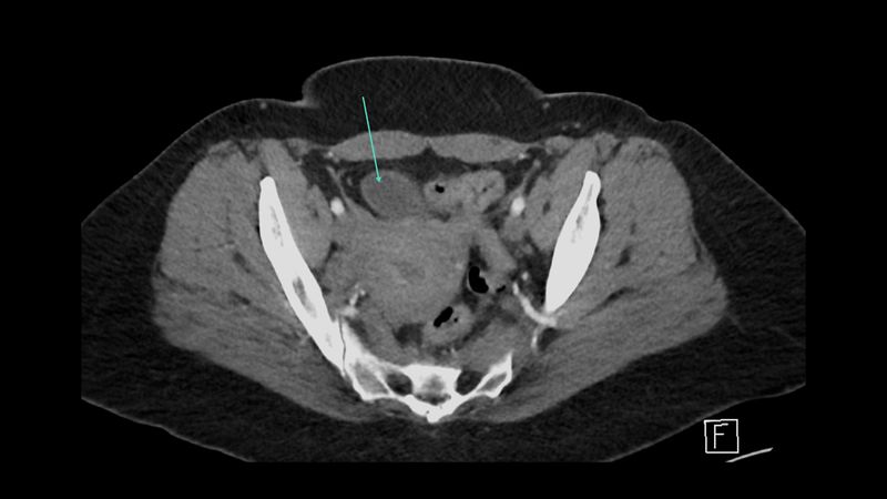
Which anatomical structure is arrowed?
Single answer — type the structure
Correct — Urinary bladder
Bony landmarks
The pelvic ring — iliac bones, sacrum, acetabula and pubic symphysis — orients every study. Read from the iliac crests down to the ischial tuberosities.
Vessels and organs
Follow the iliac vessels as they divide, then place the bladder, rectum and the sex-specific organs around them. Bowel loops fill the remaining space.
Contrast and planes
Contrast highlights the iliac vessels and organ enhancement; coronal and sagittal reformats clarify the bladder, uterus or prostate. Knowing the plane helps you name structures.
Keep practising by region
Move through the rest of cross-sectional anatomy one region at a time — brain and abdominal CT, cardiac MRI and more — and let each explanation fix the structure in memory.
Ready for the full set?
Get 20 free questions across 8 body regions — name the structure, get the explanation, and track what sticks.
Start free — 20 questions