 RadiologyQuizLab
SPINE MRI ANATOMY · SAGITTAL · AXIAL
Spine MRI anatomy
Spine MRI anatomy runs from the cervical cord to the lumbar roots. Practise naming the structures on real MRI slices — the fastest way to make spinal anatomy stick. Free, no account needed.
Try it now — read the slice, name the arrowed structure:
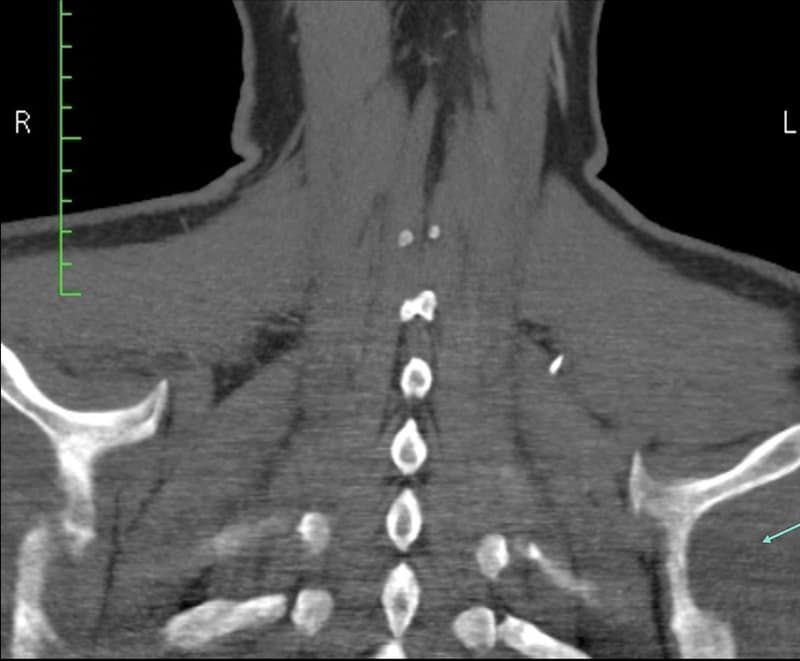
Which anatomical structure is arrowed?
Single answer — type the structure
Correct — Left infraspinatus
Vertebrae and discs
Start on the sagittal: count the vertebral bodies and the intervertebral discs, with the anterior and posterior longitudinal ligaments framing them. This is how every spine study is oriented.
Cord, conus and roots
Follow the spinal cord to the conus medullaris, then the cauda equina and exiting nerve roots. On the cervical spine, watch the cord within the canal at each level.
Canal and neural foramina
On axial images, assess the central canal, the lateral recesses and the neural foramina where the roots exit. Naming these on real images turns recognition into recall.
Keep practising by region
Move through the rest of imaging anatomy one region at a time — brain and body CT, cardiac and musculoskeletal MRI — and let each explanation fix the structure in memory.
Ready for the full set?
Get 20 free questions across 8 body regions — name the structure, get the explanation, and track what sticks.
Start free — 20 questions