 RadiologyQuizLab
SHOULDER MRI ANATOMY · OBLIQUE PLANES
Shoulder MRI anatomy
Shoulder MRI anatomy centres on the rotator cuff and the labrum, read across oblique coronal, oblique sagittal and axial planes chosen to follow the tendons rather than the body axes. The way to learn it is to name each structure on real images. This guide covers the rotator cuff, the labrum and capsule, and the biceps and bony landmarks — then lets you practise, free.
Try it now — read the slice, name the arrowed structure:
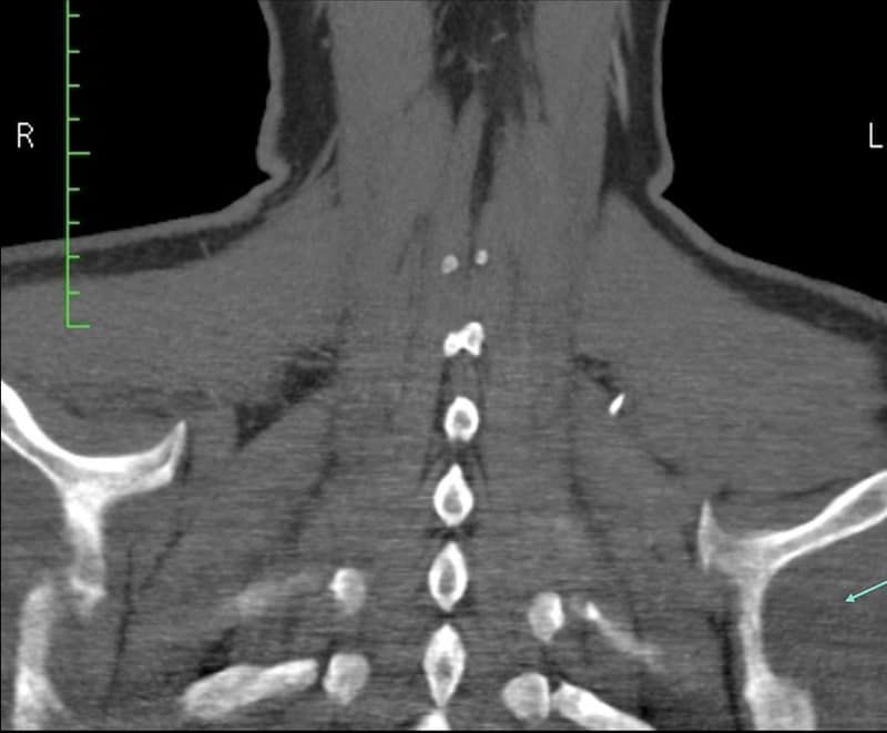
Which anatomical structure is arrowed?
Single answer — type the structure
Correct — Left infraspinatus
The rotator cuff
The four rotator-cuff muscles and tendons wrap the humeral head: supraspinatus on top, infraspinatus and teres minor behind, and subscapularis in front. On the oblique coronal plane the supraspinatus tendon is followed from its muscle belly to its insertion on the greater tuberosity — the site of most tears, and where most of your attention goes. The oblique sagittal plane shows all four tendons together in cross-section, the best view for learning how they sit around the joint.
Labrum and capsule
The glenoid labrum is a rim of fibrocartilage that deepens the socket; on axial images it appears as a dark triangle at the front and back of the glenoid, and the superior labrum blends with the origin of the long head of biceps. Around it, the joint capsule and the glenohumeral ligaments form the static stabilisers. Learning the normal shape of the labrum at each clock position is what lets you spot when something is off.
Biceps and bony landmarks
Trace the long head of the biceps tendon from the superior labrum out through the rotator interval and down the bicipital groove between the tuberosities. Orient the whole study on the bony landmarks — the humeral head, the glenoid and the acromion above — and note the acromioclavicular joint. Naming these repeatedly on real images is what turns recognition into recall.
Keep practising by region
Move through the rest of imaging anatomy one region at a time — brain and body CT, cardiac and musculoskeletal MRI — and let each explanation fix the structure in memory.
Ready for the full set?
Get 20 free questions across 8 body regions — name the structure, get the explanation, and track what sticks.
Start free — 20 questions