 RadiologyQuizLab
How to use it
- Pick a region, then a scan (liver, carotid, shoulder, knee…).
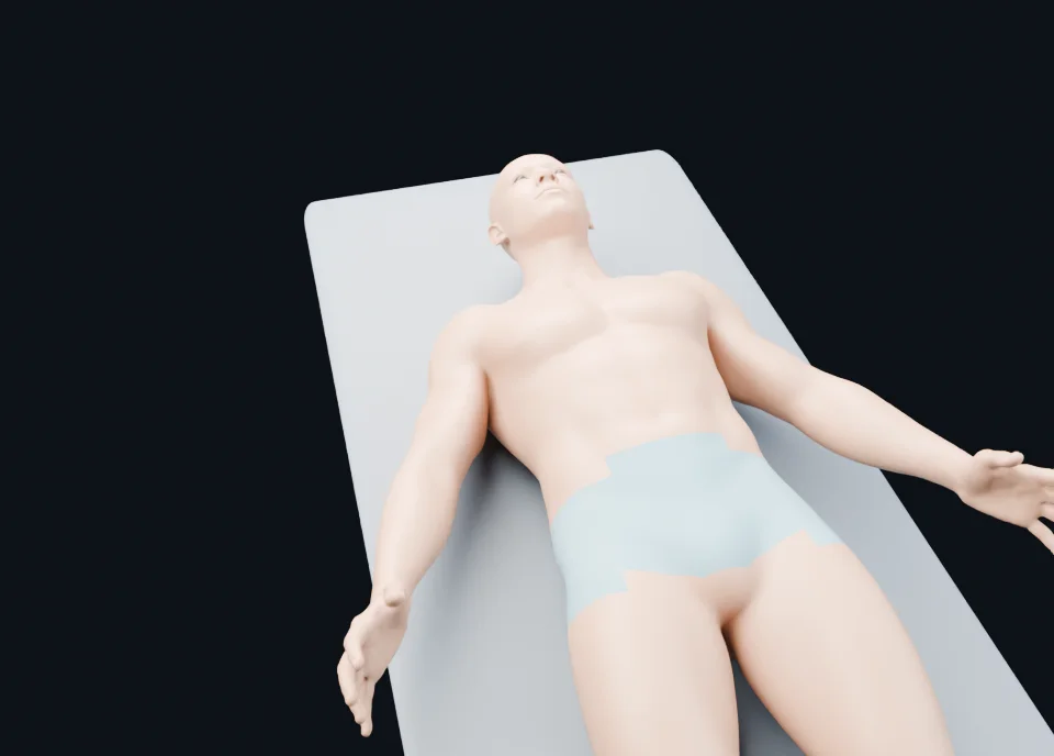
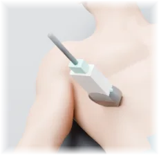
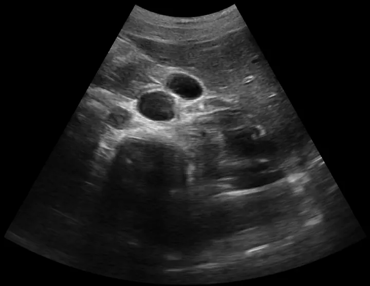
- Grab the probe on the patient and slide it along its path. You can also drag straight across the ultrasound image.
- Read the labels on the edges of the image (top = skin, left = head or proximal, depending on the plane).
- Switch to Quiz: an arrow points to a structure and you type its name, in English or in Latin.
The real gesture of each scan
Every scan reproduces the probe movement measured on the source video. For the patellar or Achilles tendon the probe slides along the limb. For the liver or the supraspinatus it stays on the same spot and fans. For the femoral and popliteal veins you press: the vein collapses, the artery stays round. We only kept clips in which the image really changes from the start to the end of the gesture.
The 32 scans
Normal anatomy only, grouped by region. The liver and the patellar tendon are free; the other scans are part of the RadiologyQuizLab subscription at no extra cost.
Abdomen
- Liver · Right subcostal view · Free
- Abdominal aorta · Longitudinal then transverse
- Appendix · Right iliac fossa, long axis then cross-section
- Left kidney · Longitudinal then transverse
- Right kidney · Longitudinal then transverse
- Hepatic veins · Right subcostal oblique view
- Spleen · Longitudinal then transverse
Pelvis
- Urinary bladder · Suprapubic transverse and longitudinal views
- Ovary · Right ovary, suprapubic transabdominal view through the bladder
- Testis · Right testis, longitudinal and transverse views
- Uterus · Suprapubic transabdominal views, longitudinal then transverse
Neck
- Common carotid and thyroid lobe, transverse sweep · Transverse view, left side of the neck
- Common carotid artery, long axis · Longitudinal view, left side of the neck
- Parotid gland · Left parotid gland, longitudinal and transverse views
- Submandibular gland · Left submandibular gland, transverse and longitudinal views
- Thyroid lobe, long axis · Longitudinal view, left thyroid lobe
Chest
- Pleural line between two ribs · Longitudinal view, upper anterior chest
Upper limb
- Anterior shoulder, transverse sweep · Transverse view, right shoulder (anterior)
- Common extensor tendon · Longitudinal view, left elbow (lateral)
- Ulnar collateral ligament · Longitudinal view, right elbow (medial)
- Median nerve at the carpal tunnel · Transverse view, right wrist (palmar side)
- Supraspinatus tendon · Longitudinal view, right shoulder
Lower limb
- Patellar tendon · Longitudinal view, right knee (anterior) · Free
- Achilles tendon · Longitudinal view, right heel (posterior)
- Anterior hip joint · Oblique sagittal view along the femoral neck, right hip
- Femoral trochlea · Transverse suprapatellar view, right knee
- Femoral vein compression · Transverse view, right thigh (femoral vessels)
- Greater trochanter · Transverse view, right hip (lateral)
- Infant hip · Infant (baby lying on its side): lateral coronal Graf view of the hip, normal type I
- Medial collateral ligament · Coronal view, medial side of the right knee
- Popliteal vein compression · Transverse view, back of the right knee
- Tibialis posterior tendon · Transverse view behind the medial malleolus, right ankle
Finding your way on the image
The top of the image is the skin under the probe; the bottom is depth. In a longitudinal view the left of the screen is the head or proximal side. In a transverse view the left of the screen is the patient's right, as on CT. These labels sit on every image and are summed up below each scan.
A typed-answer quiz, not multiple choice
In the quiz an arrow marks a structure (common carotid artery, median nerve, supraspinatus tendon…) and you type its name. Marking tolerates typos and accepts synonyms in six languages. It is the same method as the site's 1,500+ CT, MRI and X-ray questions.
Create a free accountWhere the images come from
The ultrasound clips come from the Suresult video library, released under CC BY 4.0 and credited under each image. The patient is built from Blender Human Base Meshes (public domain, CC0). Arrows and labels are reviewed by a radiologist; the tool is for learning anatomy, not for diagnosis.
Frequently asked questions
Is the ultrasound simulator free?
Yes for the liver and the patellar tendon, with no sign-up. The other 30 scans are included in the RadiologyQuizLab subscription at no extra cost.
Do I need a probe or a machine?
No. Everything runs in the browser, with a mouse or a finger on a phone.
Are these real ultrasound images?
Yes. They are real scans of normal anatomy; no image is generated.
Who is it for?
Medical students, residents, sonography and radiography students, and anyone starting point-of-care ultrasound who wants to recognise normal anatomy.
Does it include pathology?
No. The simulator shows normal anatomy only.
Keep learning
Take the quiz
Create a free account to take the quiz on the free scans and try the first 20 CT, MRI and X-ray questions.
Create a free account